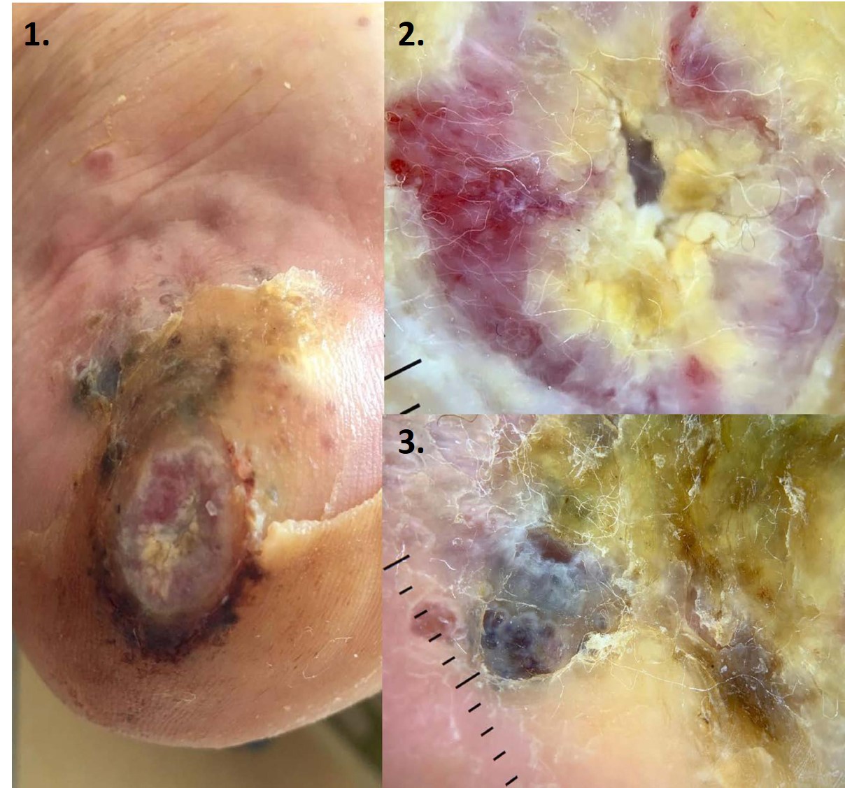
Acral Melanoma Mimicking a Non-Healing Arterial Ulcer
Image Description
Acral melanomas sums up a percentage of 2-3% (1). Acral melanomas are more common in weight-bearing areas of the foot (2), including the calcaneal zone where our patient’s melanoma was identified. Acral melanomas can adopt an amelanotic appearance, which may be attributed to a lack of melanin production and neovascularization (3). Due to their atypical clinical features and rarity, acral melanomas tend to present later and are difficult to diagnose.
We present the case of a 60-year-old female, known with hypertension and a 4-year history of a left plantar ulcer. At that time, it was thought to have been an arterial ulcer and was managed symptomatically. When presented to our dermatology department she complained of significant pain. Arterial Doppler ultrasonography detected atherosclerotic irregularities but no occlusion. After dermoscopy exam with suspicious signs (irregular fibrillar pattern showing structureless pigmentation and polymorphic vascular pattern), we performed a punch-biopsy and subsequent histology confirmed an invasive acral melanoma with 3.9 mm Breslow thickness.
Acral melanomas can present with bleeding or as a wound that fails to heal (4). This case demonstrates that persistent, non-healing foot ulcers should be evaluated for alternative diagnoses. Melanoma should be considered in cases such as this and histological evaluation performed, especially when a foot ulcer displays atypical features and/or persists despite treatment. This case underlines the importance of considering skin malignancies in cases of chronic, non-healing ulcered lesions of feet. Furthermore, we point out the dermoscopy being an important tool in the diagnosis of acral melanoma.
References
Yin NC, Miteva M, Covington DS, Romanelli P, Stojadinovic O. The importance of wound biopsy in the accurate diagnosis of acral malignant melanoma presenting as a foot ulcer. Int J Low Extrem Wounds. 2013 Dec;12((4)):289–92.
Costello CM, Pittelkow MR, Mangold AR. Acral Melanoma and Mechanical Stress on the Plantar Surface of the Foot. N Engl J Med. 2017 Jul;377((4)):395–6.
Muinonen-Martin AJ, O'Shea SJ, Newton-Bishop J. Amelanotic melanoma. BMJ. 2018 Mar;360:k826.
Pereda C, Traves V, Requena C, Serra-Guillén C, Llombart B, Sanmartín O, et al. Clinical presentation of acral lentiginous melanoma: a descriptive study. Actas Dermosifiliogr. 2013 Apr;104((3)):220–6.