Skin lesions mimicking melanoma
Image Description
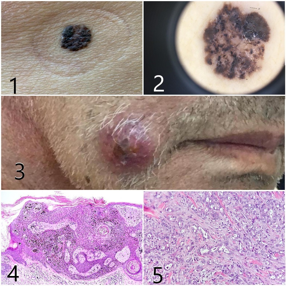
Despite the increasing awareness of melanoma over the last 40 years, clinical diagnostic accuracy remains disappointing. Benign pigmented lesions can clinically simulate melanoma (false positive)[1]. A 70 year old patient presented to us with altered general condition and jaundice, an irregular, pigmented tumor formation, with chromatic polymorphism on the surface, with achromic areas - possibly central regression, at the left flank (Figure 1; Figure 2), evolving for about 2 years and an erythematous tumor node, with central necrosis area, at the level of the right hemiface (Figure 3). An abdominopelvic CT was performed, which showed multiple round-oval formations with variable diameters (metastases), located at the level of the liver and tail of the pancreas. The biopsy of the left flank lesion showed pigmented seborrheic keratosis in collision with superficial basal cell carcinoma. From the lesion of the face a punch biopsy was performed that showed tumour proliferation in the deep dermis made up of glandular structures, beams and cords of cells with pleomorphism, indicating a cutaneous metastasis from an adenocarcinoma with visceral origin (Figure 4; Figure 5). Immunohistochemical tests were diffusely positive for CK7 and focally positive for CK20, the immunophenotypic profile indicating the pancreatic origin of the tumour process. Our case highlights the atypical basal cell carcinoma characterized by equivocal dermatoscopic features typical of malignant melanoma can be difficult to diagnose, thus secondary skin lesion has been associated with the misdiagnosis of the lesion [2]. Histologic examination is therefore important to ensure proper diagnosis [3].