Ocular Toxoplasmosis
Image Description
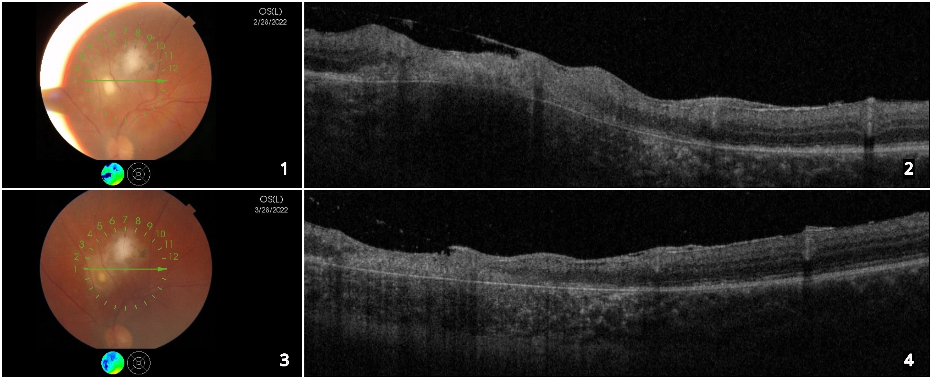
A 25-year-old male presented to our clinic with blurry vision in the left eye, for about 1 month. He had a history of ocular toxoplasmosis for which he was under care, but he stopped using his medication one week later. His best corrected visual acuity was 6/48 in the right eye and 6/24 in the left eye, on the Snellen chart. The anterior segment of both eyes was unremarkable. Fundus examination was normal in the right eye, while the left eye had vitritis and two active lesions of focal retinochoroiditis outside the temporal vascular arcade (Figure 1). OCT of the left eye showed increased thickness in the superficial layers of the retina with hyper-reflective particles in the vitreous adjacent to the retina (Figure 2). Serology was positive for Toxoplasma gondii, with detectable IgG antibodies. After 1 month of follow-up under treatment, the patient’s visual acuity was increased, the fundus examination showed clear vitreous and white inactive retinal choroiditis scars (Figure 3), and the OCT scan showed decreased thickness of the retina (Figure 4).
Ocular toxoplasmosis is one of the most common causes of infectious chorioretinitis. Depending on the retinal location, this condition may cause substantial vision impairment [1]. The treatment includes antimicrobial drugs and corticosteroids and is maintained for 4–6 weeks [2]. Recent studies revealed that the mean visual acuity significantly improves after treatment and lesion size decreases during the initial months with limited change after that [3, 4].
References
Smith J.R., Ashander L.M., Arruda S.L., Cordeiro C.A., Lie S., Rochet E., Belfort R.Jr., Furtado J.M., Pathogenesis of ocular toxoplasmosis, Prog Retin Eye Res., 2021, 81
Kalogeropoulos D, Sakkas H, Mohammed B, Vartholomatos G, Malamos K, Sreekantam S, Kanavaros P, Kalogeropoulos C. Ocular toxoplasmosis: a review of the current diagnostic and therapeutic approaches. Int Ophthalmol. 2022 Jan;42(1):295-321. doi: 10.1007/s10792-021-01994-9. Epub 2021 Aug 9. PMID: 34370174; PMCID: PMC8351587.
Ozgonul C., Besirli C.G., Recent Developments in the Diagnosis and Treatment of Ocular Toxoplasmosis, Ophthalmic Res. 2017, 57:1-12.
Vishnevskia-Dai V., Achiron A., Buhbut O., Berar O.V., Musika A.A., Elyashiv S.M., Hecht I., Chorio-retinal toxoplasmosis: treatment outcomes, lesion evolution and long-term follow-up in a single tertiary center, Int Ophthalmol 2020, 40(4): 811-821.