Foreign body at the level of rectal tumor formation
Image Description
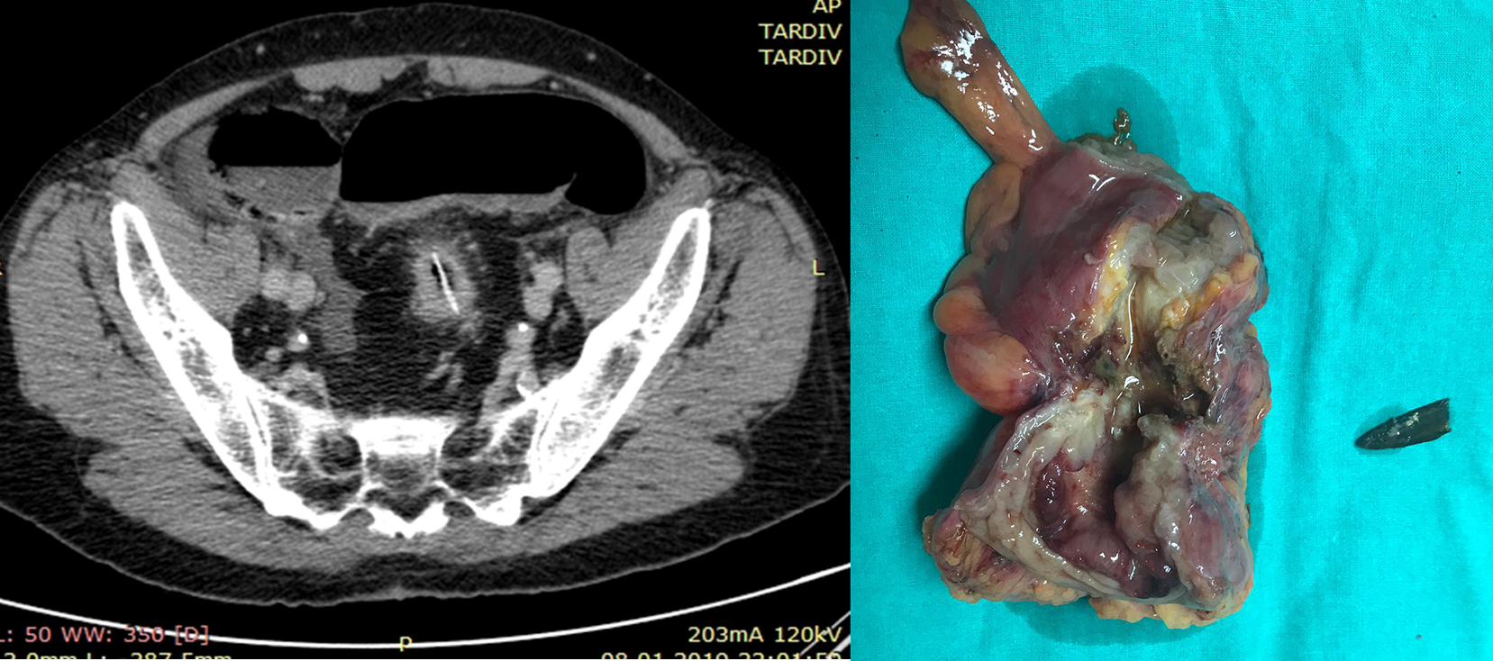
We present the case of a 75-year-old patient presenting with nausea, faecal emesis and intense abdominal meteorism, symptoms that began three before the presentation and accentuated within the last 12 hours. Clinical examination revealed an increased volume of the abdomen, painfully diffused on palpation. Abdominal radiography highlights multiple air-fluid levels. Computerized tomography: Parietal, circumferential thickening with spiked contours of the middle sigmoid, associating the important distension of the overlying sigmoid, and, at the level of the stenosis, endoluminal, a linear image of 3mm thick and 24mm in length, spontaneous hyperdensity (180UH), important densification of neighbouring fat. A rectosigmoidectomy with left flank colostomy – Hartmann’s operation, is performed. The opening of the lumen of the resected colonic portion reveals a foreign body (wooden fragment) impacted at this level. Histopathological diagnosis is colonic adenocarcinoma with a high degree of cellular differentiation (G1) invasively up to serum pT3N0. The particularity of the case consists in the difficulty of the pre- and intraoperative differential diagnosis between neoplasia requiring oncological visa management and an inflammatory pseudotumoral formation with a more limited surgical management. In an emergency, the intraoperative macroscopic examination of the resection piece is essential. Differentiation between tumour and inflammatory aspects is difficult, which is why the presumptive diagnosis and the surgical decision must be inclined to the lesion with the most severe evolutionary potential.